
Bladder pain syndromes (VDOS) are a painful, disabling and debilitating chronic condition of unknown etiology, complex diagnosis and often discouraging treatment. There are several theories that try to explain their origin and the symptoms they cause. There is no examination or analysis that allows to clearly reach its diagnosis, but in approximately 10% of the cases a characteristic lesion called Hunner’s lesion is detected by urinary endoscopy (cystoscopy) and is diagnostic of interstitial cystitis (IC).
IC is a disease included in the catalog of rare diseases. It translates a series of severe chronic inflammatory changes similar to those found in other systemic diseases such as autoimmune diseases or rheumatic diseases, but with specific involvement of the bladder that suffers severe deterioration with detachment of its coating and increased permeability. Urine is rich in toxic elements that induce inflammation and pain and make the picture chronic because they lead to increased cell destruction and the inability to induce adequate turnover by new mature cells that restore the barrier function and waterproofing.
Hunner’s lesion is only the punctual manifestation of a global bladder disease and its treatment requires the elimination of that lesion, although the risk of them appearing again is high, a circumstance that occurs in an average period of 8 months.
90% of patients with VDOS who do not have Hunner lesion do not usually show inflammatory changes as evident as in what they do have Hunner’s lesion and the pain seems related to a disorder in the permeability of the bladder and a dysfunction of the transmission of sensation in the nerves and in the central nervous system.
The treatment of these painful syndromes follows an algorithm that includes simple analgesics or morphic derivatives of progressive potency, antidepressants and antiepileptics of pain-modulating effect, instillation of different drugs into the bladder by means of a probe (hyaluronic acid, chondroitin sulfate, DMSO, corticosteroids or anesthetics), injection of botulinum toxin, electrical neuromodulation and in some cases, immunosuppressants such as cyclosporine A. In the most severe cases that do not respond to any treatment, only the removal of the diseased bladder can be resorted to.
In recent years, evidence has been obtained on the usefulness of autologous platelet-enriched plasma (PRP) and the use of stem cells in chronic degenerative and inflammatory diseases in different organs and tissues of our body.
There are studies in animal models and some studies in humans on the usefulness of platelet-enriched plasma (PRP) in the treatment of bladder pain syndrome. The results are encouraging given that a significant improvement rate of up to 70% of patients has been published after 4 intravesical injections (1 every month). This improvement was maintained in subsequent controls. The only published human study was conducted in patients without Hunner’s injury, but unpublished experience supports the theory that improvement is more significant and prolonged in patients with Hunner’s injury, where there is a severe inflammatory basis.
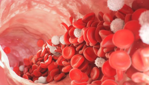
Platelets are rich in growth factors and are involved in inducing a new inflammatory process that leads to faster and more effective repair. It induces cell proliferation and migration to damaged tissue, promoting cell differentiation and maturation and angiogenesis (stimulation of new blood vessels).
It is important in urothelial repair, increasing the number of fibroblasts and smooth muscle cells, promoting oxygenation and subsequent tissue remodeling. These effects, together with other growth factors such as EGF, promote the regeneration of damaged urothelium and counteract fibrosis resulting from the disease.
PRP can also be applied to treat torpid ulcers as well as neuropathic pain because it promotes neuronal regeneration. It facilitates axonal regeneration and thus modulates aberrant pain signals coming from the bladder.
It also decreases the number of episodes of bacterial cystitis because it improves the urothelial barrier and prevents damage caused by uropathogenic bacteria
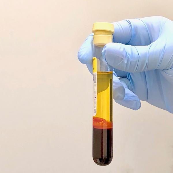
The treatment is performed in the operating room on an outpatient basis (without admission) and with sedation or intradural anesthesia. Previously, on the same day of the procedure, blood is extracted from the patient and from it his plasma is isolated and concentrated that is enriched with his own platelets. At the time of injection that plasma is chemically activated and immediately after it is injected at different points in the bladder.
This treatment is repeated a total of 4 times (1 every month) as advised by published evidence. There is some recent study (2022) that shows a significant improvement with a single treatment, but the results are clearly lower than those obtained with the repetition of the treatment, at least 3 times.
Like any invasive procedure, it has some risk of complication. However, these complications are no greater than those associated with any other minor endoscopic intervention. As a result of the introduction of an endoscopic instrument and needle puncture at various points of the bladder, transient pain, persistent voiding desire sensation, even with empty bladder, light bleeding or infection may occur. Very rarely the bleeding may be heavier and require placement of a flushed probe for a few hours. Prior to the procedure, a preventive antibiotic is administered that should continue for a few more days. There are no reactions to the injected product since it is obtained from the patient’s own blood.
Stem cell therapy in bladder pain is still under investigation and is only performed in the context of controlled clinical trials and is not currently a standard treatment option.
Dr. Eduardo Vicente Palacio
Uros Associats